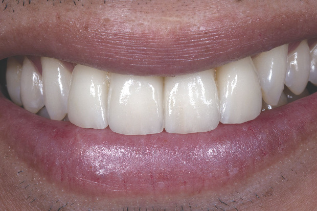
Figure 18
Close-up facial view of the final restorations.
--------------------------------------------------------------------------------------------
Seunghoon (Steve) Lee, CDT, MDC, works at Zahnwerkstatt in Beverly Hills, CA, owned by Joachim Kern, MDT. Previously, he owned a laboratory in Gaithersburg, MD until he moved to California to attend the UCLA Master Dental Ceramist Program from which he graduated in 2008, was named the Master Dental Ceramist of the Year and where he studied with Dr. Ed McLaren. Lee also lectures on IPS e.max all over the world in English and Korean. He has earned the following technical certifications: CEREC AC Scanner, Designer and Technician; CEREC inLab System Technician; E4D Designer and Technician; COS Scanner Designer; Lava 3M Designer; CAPTEK; Nobel Procera Optical Scanner Designer; and IPS e.max and Empress Instructor and Trainer.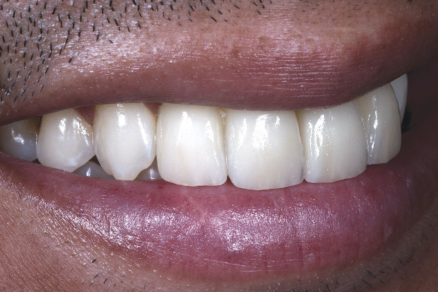
Figure 17
Close-up right lateral view of the final restorations.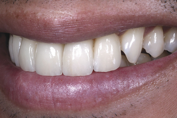
Figure 16
Close-up left lateral view of the final restorations.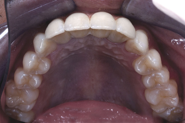
Figure 15
The restorations blend seamlessly with the patient’s surrounding natural dentition, and the artificial gingival architecture is well-defined and appears natural.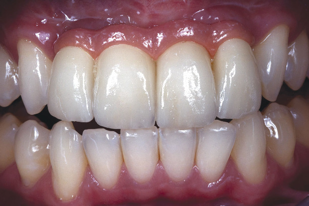
Figure 14
The framework was screw retained and then the four single IPS e.max crowns were cemented onto it. To fabricate the crowns, a full contour waxup was made, pressed with LT A1 e.max ingots and then cutback and layered with e.max porcelain.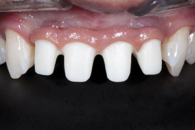
Figure 13
The gingival architecture was stained and glazed, and the entire framework was tried into the patient’s mouth to check fit, contour and esthetics.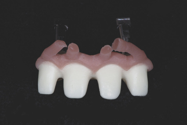
Figure 12
The IPS e.max ZirPress gingiva ingot in shade G3 was pressed onto the zirconia framework.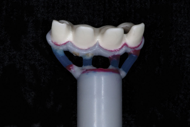
Figure 11
The waxup was sprued onto a 200-g IPS e.max ZirPress ring.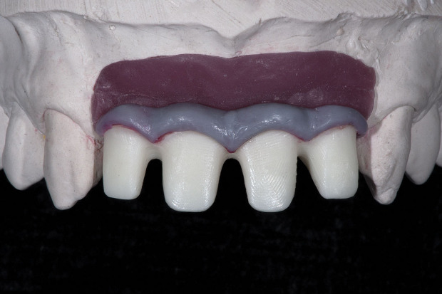
Figure 10
The gingival tissue waxup was verified on the working model.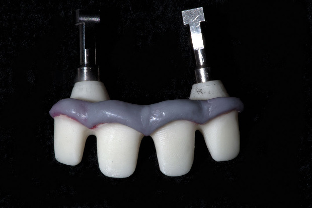
Figure 9
A waxup design of the gingival tissue to surround the restorations was added to the zirconia framework.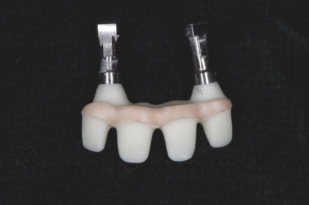
Figure 8
IPS e.max ZirLiner was applied to the zirconia framework and fired.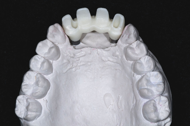
Figure 7
The zirconia framework was tried on the working model.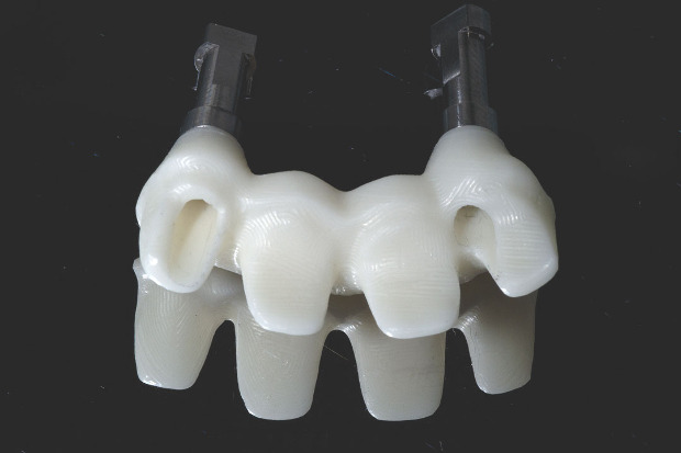
Figure 6
The lingual aspect of the zirconia framework was designed to be screw-retained through the gingival architecture to the implants.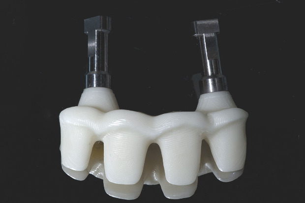
Figure 5
A four-unit zirconia framework was designed and milled using the Nobel Procera system.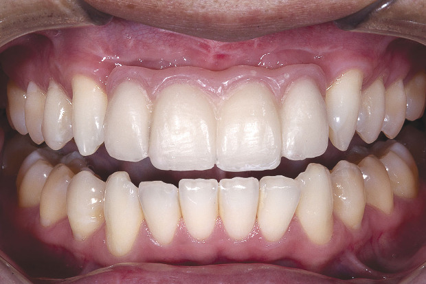
Figure 4
The patient wore this new esthetic functional prototype (designed by Dr. Edward McLaren) for about two months so he could evaluate his function, speech and different movements and see the anticipated gingival effects, natural architecture and contours that were lacking in the previous restorations. This process achieves the best restorative outcome for the patient.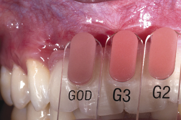
Figure 3
Shade tabs were held adjacent to the patient’s natural gingiva to determine the appropriate shade that would be used to press artificial gingiva onto the new zirconia-based framework, creating the illusion of gingival tissue. IPS e.max ZirPress ingot G3 was selected.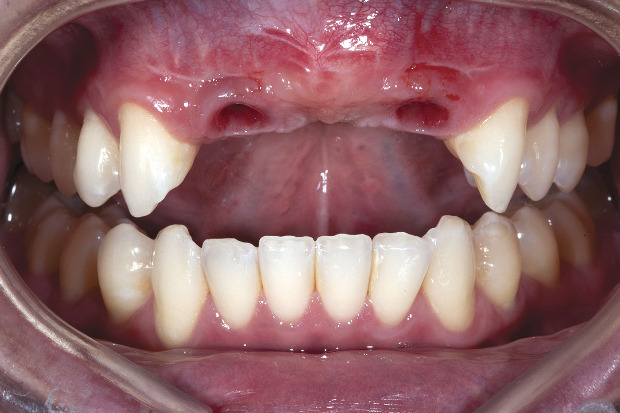
Figure 2
The restorations were removed and loss of gingival architecture was noted. There was no curved or natural shape to the gingival margins and no papilla tissue to create a natural appearance. Additionally, one of the implants sat between the #7 and #8 sites.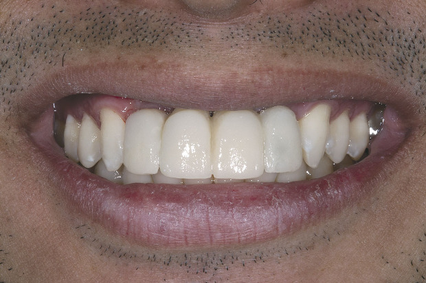
Figure 1
The patient’s maxillary incisors were implant-supported restorations that were unnaturally long, white and opaque. The restorations would be replaced to achieve a more esthetic and natural appearance.

Article
The challenging aspect of this case was that one of the implants was improperly placed between #7 and #8. Steve Lee explains how he made a screw-retained zirconia framework with pink gingiva tissue to recreate the lost gingiva and then cemented on four...

Author
Steve Lee
Master Dental Technician at Zahnwerkstatt USA · Los Angeles, CA
Until November 2012, Professional instructor / Co-director of the UCLA Master Dental Ceramist program. Now, working as a Master dental technician in Beverly Hills, Zahnwerkstatt, LLC U.S.A on North Camden Drive.
Subscribe to LMT Magazine. It's FREE to U.S. addresses.


September 2014
Browse all articles from the September 2014 issue of LMT Magazine
-

IPS e.max ZirPress
Ivoclar · Consumables
-

Implants
4,041 subscribes


-

Ivoclar
Company
-

Ed McLaren
CEO at Artoral America · Park City, UT
-

Herve Gross
Dental Technician at Panthera Dental · Quebec, QC
-
Smart Redesign Delivers More Space in Same Footprint
Kim Molinaro · Inspiring Interiors · June/July 2025
Instead of building a costly addition to its Maumee, OH, facility, Lantz Dental Prosthetics worked with an architect to create a better layout within its...
Here are eight strategies for ensuring a safer environment for your business.
Renstrom Dental Studio: Truly a Family
Jennifer Ludwig · I Want to Work in That Lab · June/July 2025
Lino Lakes, MN; 72 employeesOwners: Randi and Scott James A third-generation, family-run business, Renstrom Dental Studio in Lino Lakes, MN, is built...
Cosmetic Dental Specialties: Quality Over Quantity
Jessie Blanchard · I Want to Work in That Lab · June/July 2025
Portland, OR; 15 employeesOwner: Brianne Lequerica-Munday When I first joined Cosmetic Dental Specialties in Portland, OR, in 2021, I was used to being...
Trains, Planes and Automobiles: The Global Impact of 3D Printing
Kelly Fessel Carr · Publisher's Page · June/July 2025
As we were organizing our Product Focus on 3D Printing Materials for this issue